Cryo Electron Microscopy
Prof. Dr. Rasmus Schröder
We specialize in advancing Cryo Electron Microscopy (Cryo-EM), a revolutionary technique now established as a routine application on campus with a network of four dedicated microscopes.
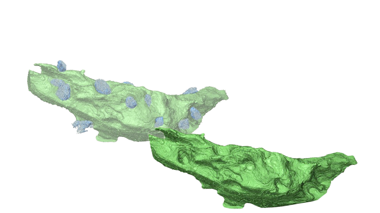
In line with Heidelberg's "Engineering Molecular Systems," we've expanded into Volume and Analytical Electron Microscopy, essential for the "3D Matter Made to Order" Excellence Cluster. Our group focuses on sample preparation and imaging techniques for hybrid systems, combining biological and man-made materials, bridging life sciences and engineering to study structure-function relationships.
Research Strategy
The last years saw a revolution in Cryo Electron Microscopy. One focus of our work was to establish this structural biology technique as a routine application for the wider campus. A network of four dedicated microscopes is now installed and provides a platform for many groups on campus. However, scientific interests in the framework of Heidelberg’s new activity “Engineering Molecular Systems” asked for further application development of Volume Electron Microscopy and also new ways of Analytical Electron Microscopy of carbon-based materials. These activities were paramount for establishing a strong structural focus of Heidelberg’s part in the Cluster of Excellence “3D Matter Made 2 Order”. In this environment, our group is studying sample preparation and imaging techniques for hybrid systems, i.e., engineered systems combining molecules, cells, and tissue with man-made carbon materials. With these activities, we are building a strong bridge between life sciences and the emerging engineering sciences on campus for studying structure-function relationships.

Our microscopies have advanced to such an extent, that we can now move from studying simple systems, e.g., reconstituted actomyosin systems, to more complex and hybrid systems. Examples are sarcomeric structures, where we want to shed light on the details of the initial interaction of myosin with cardiac thin filaments to understand how this can lead to cardiomyopathies. Here, the combination with engineering approaches, such as growing heart muscle cells in 3D-printed, active scaffolds, will be explored. These studies will also need to merge morphological volume microscopy with the detailed analysis of molecular structures in cryo-electron tomography. Furthermore, we will combine this work with our developments of analytical microscopy, testing a new approach to correlate light and electron microscopy by transferring spectroscopic techniques from fluorescent inorganic materials to organic fluorophores. Preliminary results indicate, that we no longer destroy organic molecules when imaging with ultra-low energy electrons – allowing a truly molecular correlation between light and electron microscopy.
Project Leader

Prof. Dr. Rasmus Schröder
- +49 (0)6221 54-51350
- rasmus.schroeder@bioquant.uni-heidelberg.de
Selected Publications
Two-step absorption instead of two-photon absorption in 3D nanoprinting
Vincent Hahn, Tobias Messer, N. Maximilian Bojanowski, Ernest Ronald Curticean, Irene Wacker, Rasmus R. Schröder, Eva Blasco & Martin Wegener
Nature Photonics volume 15, pages 932-938 (2021)
Thermoresponsive Hydrogels with Improved Actuation Function by Interconnected Microchannels
Tobias Spratte, Christine Arndt, Irene Wacker, Margarethe Hauck, Rainer Adelung, Rasmus R. Schröder, Fabian Schütt, Christine Selhuber-Unkel
Adv. Intell. Syst., 4: 2100081.(2021)